Phage structure captured for the first time, to benefit biotech applications
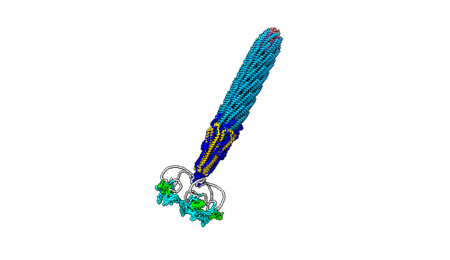
Phage image based on that published in Gold et al, Nature Communications
New insights into the structure of phages will enable researchers to develop new uses for the viruses in biotechnology.
Phages are viruses that infect bacteria, which enables them to be exploited as tools in biotechnology and medicine. Now, for the first time, researchers at the University of Exeter, in collaboration with Massey University and Nanophage Technologies, New Zealand, have mapped out what a commonly-used form of phage looks like, which will help researchers design better uses in future.
One common use for phage is phage display, which is a useful tool in drug discovery. Phage display works by linking a gene fragment of interest to a phage gene that makes one of the phage coat proteins. The new coat protein with the linked protein of interest appears on the surface of the phage, where it can be assayed and tested for biological activity.
Billions of types of phages exist. Phage display often uses a type of phage known as filamentous, so called because they are long and thin, making the display of many proteins across its surface possible. Although phage display and other applications have proved successful, until now, scientists have not known what this type of phage looks like.
For the first time, Dr Vicki Gold at the University of Exeter, has revealed the structure of a filamentous phage, in research published in the journal Nature Communications. She said: “Phages form part of a very exciting and growing area of research, with a range of current and potential applications. Yet until now, we’ve not had a complete picture of what filamentous phages look like. We’ve now provided the first view, and understanding this will help us improve applications for phage into the future.”
Because filamentous phages are so long, scientists have previously failed to capture an image of their entirety. To image the phage, researchers created smaller versions, which are around 10-fold shorter, which look like straight nanorods rather than entangled spaghetti-like filaments. This mini version was small enough to be imaged in its entirety using high-resolution cryo-electron microscopy.
The paper is entitled “Cryo-electron microscopy of the f1 filamentous phage reveals insights into viral infection and assembly”, published in the journal Nature Communications. The work was funded by Wellcome.